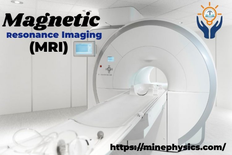
Magnetic Resonance Imaging (MRI) Scanner
MRI is a medical imaging technique that captures images of internal body structures. It is safe for soft tissues but potentially affects objects.

Definition:
Magnetic resonance imaging is a harmless medical imaging procedure that generates comprehensive images of almost every internal structure in the human body, including organs, bones, muscles, and blood vessels. A powerful magnet and radio waves are used in MRI scanners to create photographs of the body.
Invention:
Paul S. Lauterbur is credited with developing the first method of MR imaging. He used magnetic field gradients to encode spatial information into an NMR signal in September 1971 and presented the underlying theory in March 1973.
Who first developed it?
The first images were taken in the early 1970s and the first living person was taken in 1977. These machines came on the market in the 1980s and are now widely used to view the body's internal structures, particularly in soft tissues such as the brain.
Working:
When a patient is placed inside the machine. Protons in the body are compelled to align with the magnetic field produced by an MRI's powerful magnets. Then, when an RF current is passed through the patient, the protons are stimulated and unbalanced as they fight the pull of the B-field. MRI sensors can pick up the energy produced when protons rearrange with the magnetic field when the RF field is switched off. The time it takes protons to restore the B-field, as well as the amount of energy released, varies depending on the environment and the chemical nature of the molecules. Based on these properties, doctors can distinguish between different tissue types. The patient is positioned inside a sizable magnet to produce an image and must stay still throughout the imaging procedure to prevent image blurring. Contrast agents (often containing the element gadolinium) can be administered intravenously to a patient before or during a scan to increase the rate at which protons are targeted in the magnetic field. The image becomes brighter with a faster proton rearrangement.
Uses:
- MRI scanners are very well suited for examining soft tissue or non-bony bodily parts. They are different from computed tomography (CT) scans in that no dangerous ionizing X-rays are used. It can visualize knee and shoulder injuries because the brain, spinal cord, and nerves, as well as muscles, ligaments, and tendons, are visible far more clearly than on standard X-rays and CT scans.
- Aneurysms and tumors can also be identified with MRIs, which can discriminate between white and grey matter in the brain. When frequent imaging is required for diagnosis or treatment, particularly in the brain, MRI is the imaging modality of choice because it uses no radiation (X-rays or otherwise), and is hence non-invasive. A CT scan or X-ray is less expensive than an MRI, though.
- The term "functional magnetic resonance imaging" refers to one kind of specialized MRI. It is used to keep track of brain structures and identify the regions of the brain that get "activated" (consume more oxygen) when doing different types of cognitive tasks. It is utilized to deepen our understanding of how the brain is organized and offers a potential new benchmark for evaluating neurological state and neurosurgical risks.
Are there any Risks?
Even though X-rays and CT scans use ionizing radiation to produce their images, MRIs nevertheless employ a powerful magnetic field. A wheelchair might be thrown across the room by the B-field, which is strong enough to affect iron, some steel, and other magnetizable items quite strongly. The magnetic field spreads beyond the machine. Before getting a scan, patients should tell their doctor whether they are taking any medications or have any implants.
Precautions:
No one with iron-containing implants, including pacemakers, vagus nerve stimulators, implanted cardioverter defibrillators, loop recorders, insulin pumps, cochlear implants, deep brain stimulators, and capsule endoscopy capsules, is allowed to use the MRI machine.
Noise: Special hearing protection may be necessary when using some MRI scanners because of loud noises including clicks and beeps and noise intensities of up to 120 dB.
Nerve Stimulation: When the field on an MRI changes quickly, it can occasionally cause a trembling sensation.
Contrast agents - Patients with severe renal failure who require dialysis may develop nephrogenic systemic fibrosis, an uncommon but serious condition that may be linked to the use of several gadolinium-containing medicines, including gadolinium and others. Although a cause-and-effect relationship has not been proven, current American recommendations advise that gadolinium be administered to dialysis patients only when necessary and that the procedure be carried out as soon as feasible after the scan to help the body swiftly eliminate the substance.
Pregnancy: Although no effects on the baby have been proven, it is advised that pregnant women avoid having MRIs as a precaution, especially during the first trimester when the fetal organs are developing and contrast agents if employed, may enter the fetal blood.
Claustrophobia -It could be challenging for anyone who is even mildly claustrophobic to use the equipment for extended periods of time. Patients are given tools to manage pain through familiarity with the procedure and device, imaging methods, sedation, and anesthesia. Other coping strategies include covering or squishing your eyes, pressing the panic button, viewing a film or movie, or listening to music. An open MRI is a device that is open on all sides rather than a tube that is closed on one side to partially enclose the subject. It is made to accommodate individuals who dislike the constricting tunnel and distracting noises of conventional MRI as well as those whose size or weight makes traditional MRI unsuitable.
What is an Open MRI?
An imaging device that takes pictures is referred to as an open (or "open") MRI. Two flat magnets are often positioned above and below you in an open MRI machine, with enough room between them for you to lie down. This opens up the sides and significantly lessens the claustrophobia that many individuals encounter when using closed MRI scanners. Open MRIs do not, however, generate images as clearly as closed-hole MRIs. For imaging, you must lie down inside an open hole or tube created by a ring of magnets on a closed-hole MRI machine. The head-to-ceiling space in closed-hole MRI scans is constrained. Even though some people may feel anxious and uncomfortable as a result, these MRI machines deliver the best possible images. Speak with your doctor if you're anxious about getting an MRI or if you're afraid of small places. Your doctor will go over the possibilities for sedatives (drugs that help you relax) or perhaps anesthesia if necessary.
What is an MRI with Contrast?
A contrast agent may be injected during some MRI tests. Gadolinium is a rare earth metal found in the contrast material. When this chemical is inside of you, it alters the magnetic characteristics of the water molecules in the area, which enhances the clarity of your images. By doing this, diagnostic images are more sensitive and specific.
Visibility of the following is enhanced by contrast material:
Tumors.
Inflammation.
Infection.
various organs have blood flow.
a blood vessel.
Your doctor will insert an IV (intravenous catheter) into a vein in your arm or forearm if the contrast material needed for your MRI scan needs to be administered. Injecting the contrast agent will be done using this IV. Prepayments for contrast are secure. major reactions are extremely rare, however mild to major adverse effects can happen.
What is the difference between MRI and CT Scan?
- Computed tomography (CT) combines X-rays and computers to produce images of the inside of the body, whereas magnetic resonance imaging uses magnets, radio waves, and a computer.
- When examining soft tissue or non-bony bodily parts, medical practitioners frequently opt to use an MRI scan rather than a CT scan. The absence of dangerous ionizing X-rays makes MRI safer as well.
- In comparison to standard X-rays and CT scans, MRIs also produce images of the brain, spinal cord, nerves, muscles, ligaments, and tendons that are significantly clearer.
- But not everyone is eligible for an MRI. Its magnetic field can dislodge metal implants or cause pacemakers and insulin pumps to malfunction. If so, the next-best option is a CT scan.
- In general, MRI scans cost more than CT or X-ray scans.
What does an MRI show?
The inside of the body can be seen in great detail thanks to magnetic resonance imaging (MRI). With the aid of an MRI, medical experts can "view" and assess a number of different body structures, such as:
your nervous system and its surroundings.
organs of the chest and abdomen, such as the adrenal glands, kidneys, spleen, intestines, liver, and bile ducts.
breast tissues.
the spinal cord and the skeleton.
pelvic organs, such as the bladder and reproductive organs (the prostate in males and the uterus and ovaries in females) are assigned at birth.
the blood vessel.
lymph nodes.
What's Your Reaction?


