X-ray diffractometer, invention, working, applications
X-ray diffractometer, invention, working, applications
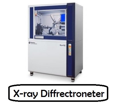
What is X-ray diffractometer?
It is a scientific instrument used to analyze the crystalline structure of materials. The structure of materials can be examined and measured using this device. It uses a technique called X-ray diffraction, which works on the principle that when X-rays collide with a crystalline material, they scatter in a pattern determined by the arrangement of atoms in the crystal lattice.
Invention:
The invention of the X-ray diffractometer cannot be attributed to one person. It is the culmination of advances made by various scientists and researchers over time.
The pioneering work on X-ray diffraction was done by Max von Laue, a German physicist who showed in 1912 that X-rays can be refracted by crystals. This discovery earned him the Nobel Prize in Physics in 1914. Von Laue's work laid the foundation for understanding X-ray diffraction as a powerful tool for studying crystalline structures.
Following von Laue's discovery, British physicists William Henry Bragg and his son William Lawrence Bragg developed Bragg's law of X-ray diffraction in 1912. This law describes the relationship between the angles of incidence and diffraction of X-rays on a crystal. lattice and allows you to determine the crystal structure.
In the early 20th century, several researchers and instrument makers contributed to the development of X-ray diffractometers. The design and implementation of diffractometers involved the integration of X-ray sources, sample holders, detectors, and goniometers.
Notable contributions were made by William Lawrence Bragg, who further improved X-ray diffraction techniques and tools. He and his collaborators developed the first practical X-ray spectrometer in 1913, which can be considered an early form of diffractometer.
Components:
The main components of an X-ray diffractometer include:
1)X-ray source: Usually a sealed tube or rotating anode X-ray generator that produces a beam of X-rays at a specific wavelength.
2)Monochromator: A device that selects a specific wavelength of X-rays from a beam generated by an X-ray source. This ensures that the X-rays used for diffraction have a well-defined wavelength.
3)Sample holder: A platform on which the sample is securely mounted and accurately positioned for analysis. The sample is usually a small crystalline material or powder sample.
4)Detector: A device that captures and measures the intensity of X-rays deflected by a sample. Commonly used detectors are scintillation counters, proportional counters or solid-state detectors.
5)Protractor: A mechanism that rotates and precisely aligns the sample and detector with respect to the X-ray beam. This rotation helps collect diffraction data from different angles.
6)Data acquisition system: collects and processes detector signals and converts them into diffraction patterns or intensity data.
Working:
An X-ray diffractometer works by directing a beam of X-rays at a crystalline sample and analyzing the diffraction pattern that occurs when X-rays interact with the crystal lattice. Here's a step-by-step explanation of how an X-ray diffractometer works:
X-ray generation: The diffractometer starts generating X-rays using an X-ray source. The most common sources are X-ray tubes or rotating anode X-ray generators. These sources produce a collimated beam of X-rays with a specific wavelength.
Monochromatic: An X-ray can contain a range of wavelengths. To obtain a well-defined wavelength for diffraction, a monochromator is used. The monochromator filters the X-ray beam by selecting a specific wavelength or a narrow range of wavelengths.
Sample preparation: Prepare a sample for analysis. In most cases, the sample is a crystalline material or some form of powdered crystals. The sample is securely attached to the sample holder, ensuring accurate placement.
Interaction of X-ray and sample: The X-ray is directed at the sample, causing the X-rays to interact with the crystal lattice of the sample. When X-rays pass through a crystal lattice, they are scattered or deflected by the atoms in the lattice.
Diffraction Pattern: Diffracted X-rays form a distinct pattern due to their interaction with the crystal lattice. This model has intensity peaks that represent certain atomic arrangements in the crystal.
Detection: A detector is placed against the sample to collect the deflected X-rays. Different types of detectors can be used, such as scintillation counters, proportional counters or solid-state detectors. The detector measures the intensity of X-rays that are deflected from different angles.
Protractor Rotation: The sample and detector are mounted on the protractor for precise rotation and alignment. The protractor rotates the sample and detector together, allowing for data collection from different angles.
Data acquisition: As the sample and detector rotate, deflected X-rays are captured by the detector and their intensity recorded. The data acquisition process involves taking measurements at specific angles to cover a range of diffraction angles.
Data Analysis: Once diffraction data is collected, it is analyzed. The data is processed and converted into a diffraction pattern or a plot of intensity versus diffraction angle. This data can be further analyzed to determine crystal structure, lattice parameters and other material properties.
Interpretation: The final step involves interpreting the diffraction pattern to extract information about the arrangement of atoms and the properties of the crystalline material. This may include comparing experimental data with known crystal structures or performing calculations to determine crystal parameters.
X-ray diffractometers are universal devices and are used in materials science, chemistry, geology, biology and other fields. They provide valuable information about the atomic and molecular structure of crystalline materials and aid in the characterization, identification and analysis of various substances.
XRD application:
Sample purity measurement.
Determine the size of the unit cell.
Characterization of crystalline materials and determination of structural properties, including:
Lattice parameters - Deformation - Grain size - Epitaxy - Phase composition - Preferred orientation.
Characterize thin film samples and measure the thickness of thin films and multilayer materials.
Determine the arrangement of the atoms.
Identification of fine-grained minerals such as clays and mixed clay layers that are difficult to detect optically.
Determination of modal amounts of minerals.
What's Your Reaction?