Microscope, principle, invention, parts, uses
Microscope, principle, invention, parts, uses
Microscope, principle, invention, parts, uses
What is microscope?
A microscope is a scientific instrument used to magnify and view small objects or details that would otherwise not be visible to the naked eye. A microscope is usually made up of several main components that work together to magnify and view microscopic objects.
What does "microscope" means?
The word "microscope" comes from the Latin "microscopium", which comes from the Greek words "micros" meaning "small" and "scopeine" meaning "to look".
Principle:
The operating principle of a simple microscope is that when a sample is brought into the focus of the microscope, a virtual, erect, magnified image is obtained at the shortest distance of clear view from the eye held on the lens.
Who invented microscope?
It is not known exactly who invented the microscope. However, the first microscopes seem to have been made around 1590 by the Dutch opticians Hans Janssen and his son Zacharias Janssen, and the Dutch instrument maker Hans Lippershey (who also invented the telescope).
History of microscope:
The history of the microscope spans several centuries and includes many inventors and achievements. Here is a brief overview of its development:
Early predecessors:
In ancient times, around 1000 BC, Egyptians used polished crystal lenses to magnify small objects.
The ancient Romans also had simple magnifying glasses, known as "burning glasses", that were used to concentrate sunlight and make fire.
In the 13th century, the Arab scholar Alhazen (Ibn al-Haytham) wrote extensively on optics and described the principles of magnification.
Invention of the compound microscope:
At the end of the 16th century, two Dutch spectacle makers, Hans Lippershey and Zacharias Janssen, are often regarded as the inventors of the compound microscope. They combined several lenses in one tube, increasing the magnification.
Their invention was developed by other Dutch scientists, including Anthony van Leeuwenhoek, who is considered the father of microbiology. Leeuwenhoek's handmade microscopes achieved great magnification and enabled him to observe microorganisms for the first time.
Refinements and improvements:
In the 17th century, the microscope underwent significant improvements. Robert Hooke, an English scientist, published the book Micrography in 1665, in which he described his observations using a compound microscope. Hooke introduced the term "cell" to the study of plant tissue.
Italian biologist Marcello Malpighi used microscopes to study the structure of plant and animal tissues, making important contributions to histology.
In the 18th century, advancements in lens grinding and polishing enabled the production of higher quality lenses, leading to improvements in microscopes.
Development of the electron microscope:
In the late 19th and early 20th centuries, the electron microscope was developed, which uses a beam of electrons instead of light to create images.
Ernst Ruska, a German physicist, designed the first electron microscope in 1931. He was awarded the Nobel Prize in Physics in 1986 for his contributions.
The electron microscope opened up new possibilities to observe even finer structures and offered greater resolution than light microscopes.
Modern Performance:
In the second half of the 20th century, technological progress led to the development of different types of microscopes, such as fluorescence microscopes, confocal microscopes and scanning electron microscopes.
These state-of-the-art microscopes offer a range of imaging modalities, including phase contrast, differential interference contrast (DIC), and laser scanning, enabling researchers to study biological samples in detail.
The microscope has played a vital role in scientific discoveries, from the exploration of the microscopic world to the development of fields such as biology, medicine and materials science. The continuous evolution and integration with other methods contributes to the continuous advancement of scientific research.
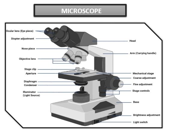
Main parts of microscope:
The specific parts may vary depending on the type of microscope, but here are the main parts of most microscopes:
Objective Lens: The objective lens is the primary lens responsible for magnifying the specimen. It is located on the bottom of the microscope and usually comes in a variety of magnifications.
Eyepiece or eyepiece lens: An eyepiece is a lens through which you look to see a magnified image. It is located at the top of the microscope and usually provides further magnification of the image formed by the objective lens.
Tube: The tube connects the eyepiece to the objective and ensures proper alignment of the optical path.
Rotating Nosepiece: The rotating nosepiece holds the objective lens and allows it to be rotated easily. This allows the user to switch between magnifications without disturbing focus or alignment.
Stage: The stage is a flat platform on which a specimen or slide is placed for observation. It usually includes a mechanism to move the slide horizontally or vertically to accurately position the specimen.
Condenser: The condenser is located under the stage and helps to focus the light on the sample. Contains lenses that collect and focus light, improving illumination and image resolution.
Diaphragm: The diaphragm is a control mechanism located in the condenser that regulates the amount of light passing through the sample. By adjusting the iris, you can control the brightness and contrast of the image.
Light source: microscopes need a light source to illuminate the specimen. Traditional microscopes use a built-in light source, such as a halogen or LED lamp, located below the stage. However, electron microscopes use electron beams instead of light.
Focusing: Microscopes have mechanisms to focus the image. The coarse focus knobs are used for the initial coarse focus, while the fine focus knobs allow you to make fine adjustments for a sharp image.
Body or Frame: The body or frame of the microscope contains and supports all components. Provides stability and allows you to properly adjust the optical system.
These are the main components of a basic microscope. However, more advanced microscopes such as electron microscopes or confocal microscopes may have technology-specific add-ons.
Types of microscope:
There are different types of microscopes, each designed for specific applications and imaging techniques. Here are some of the most commonly used types of microscopes:
1)Optical Microscope:
Compound Microscope: This is the most common type of microscope that uses visible light and a series of lenses to magnify samples. It is used for general laboratory observations and can magnify up to 2000x.
Stereo microscope: Also known as a dissecting microscope, it provides a three-dimensional view of larger, opaque specimens. It is often used to open, create, and test activities.
2)Electronic Microscope:
Transmission electron microscope (TEM): Uses a beam of electrons to pass through an ultra-thin sample and produce high-resolution two-dimensional images. TEM is suitable for studying the internal structure of cells and analyzing nanoscale details.
Scanning Electron Microscope (SEM): Scans the surface of a sample with a focused electron beam, producing detailed, high-resolution 3D images. SEM is ideal for studying surface morphology and sample topography.
3)Scanning Probe Microscope:
Atomic Force Microscope (AFM): Uses a small probe that scans the surface of the sample and measures the forces between the probe and the sample. AFM provides high-resolution images and enables you to study the surface properties of materials at the atomic and molecular level.
Scanning Tunneling Microscope (STM): It works based on the quantum tunnel phenomenon and scans the surface of conductive materials with a sharp probe. STM can provide atomic-scale resolution and is often used in nanotechnology research.
4)Confocal Microscope:
Laser Scanning Confocal Microscope (CLSM): Uses a laser beam to illuminate the specimen and collects the emitted light in a specific focal plane, excluding blurred light. CLSM provides high-resolution images and is useful for studying fluorescently labeled samples.
5)Fluorescence Microscope:
Fluorescence microscope: uses specific wavelengths of light to excite fluorescently labeled samples, allowing the visualization of specific molecules or structures in cells or tissues. It is widely used in biological and biomedical research.
6)Phase contrast microscope:
Phase Contrast Microscope: Enhances the contrast of clear and unstained samples by converting refractive index differences into visible variations. This technique is useful for observing live cells without staining.
These are just a few examples of the many types of microscopes available, but there are other specialized microscopes designed for specific purposes, such as polarizing microscopes, dark field microscopes, and digital microscopes. Each type has its own strengths and uses, allowing scientists and researchers to investigate different patterns and phenomena in different ways.
Application/Uses:
We are aware that the primary purpose of a microscope is to view items that are too small to be seen with the human eye. There are different uses for this device depending on the areas where it is used. It can be used to solve crimes, cure diseases, create new materials and even analyze fossils that are part of history.
Some application and uses of the microscope are in:
Botanical field
Biological field
Crime investigation
Educational area
Medical field
Materials science and engineering
Environmental Science
Forensic
Quality control and production
1)Microscopes in the botanical field:
This device is used by lab technicians and students who want to study the characteristics of leaves, their cells and many characteristics of a plant. Botanists conduct numerous studies on various plants and mushrooms for research purposes, which allows them to discover many new properties.
2)Biological microscopes:
We've seen this device in every biological lab. This device is used to observe microorganisms and their characteristics. In this field, microscopes are used to study bacteria, cells, and more. This device helps biologists study living organisms and their cellular structures.
3)Microscope in solving crimes:
The use of microscopes in forensic science helps simplify complex evidence and helps examine it to solve cases. We use this device for forensic purposes and prove whether the convict is innocent or not. In this field there is a great application of these devices, and without them it will be impossible to investigate some things that are not visible to the naked eye.
4)Microscopes in education:
In different institutions, colleges, schools and universities, among different optical instruments, this laboratory instrument can be found in every laboratory of major departments. Students use this device to learn new things and understand the world around them. Thanks to its excellent use, it is one of the favorite devices of students.
5)Microscopes in the field of medicine:
Man's greatest contribution to health would not have been possible without the use of microscopes. Scientists and lab technicians use this device to study various viruses and bacteria and find cures for various diseases. Researchers use this device to study deadly microorganisms and their effects on the human body.
6)Materials science and engineering:
Surface Analysis: Microscopes such as scanning electron microscopes (SEM) and atomic force microscopes (AFM) are used to study the surface morphology, structure, and composition of materials at the micro and nanoscale.
Error analysis: Microscopes help determine the causes of material defects, defects and cracks by examining the microstructure and composition of samples.
semiconductor industry. Microscopes play a vital role in the verification and characterization of integrated circuits, semiconductor devices and nanostructures used in electronics.
7)Environmental Science:
Water and Soil Analysis: Microscopes help examine microorganisms, algae, and contaminants in water and soil samples.
Pollution Monitoring: Microscopes help identify and analyze microscopic organisms as indicator species and bio-indicators to monitor environmental pollution levels.
8)Forensic:
Forensic Analysis: Microscopes are used to examine traces such as fibers, hair, pollen, and fingerprints, which can yield valuable information in criminal investigations.
9)Quality control and production:
Industrial Inspection: Microscopes aid in quality control processes by examining the structure, defects, and integrity of components, materials, and manufactured products.
Microelectronics and semiconductor industry: Microscopes are used to inspect and analyze integrated circuits, printed circuit boards and electronic components during production and testing.
Above were several applications of microscopes.
What's Your Reaction?